Allergy & Food Intolerance
Diagnostics

Allergia ELISA kit for detection of specific IgE

Test principle
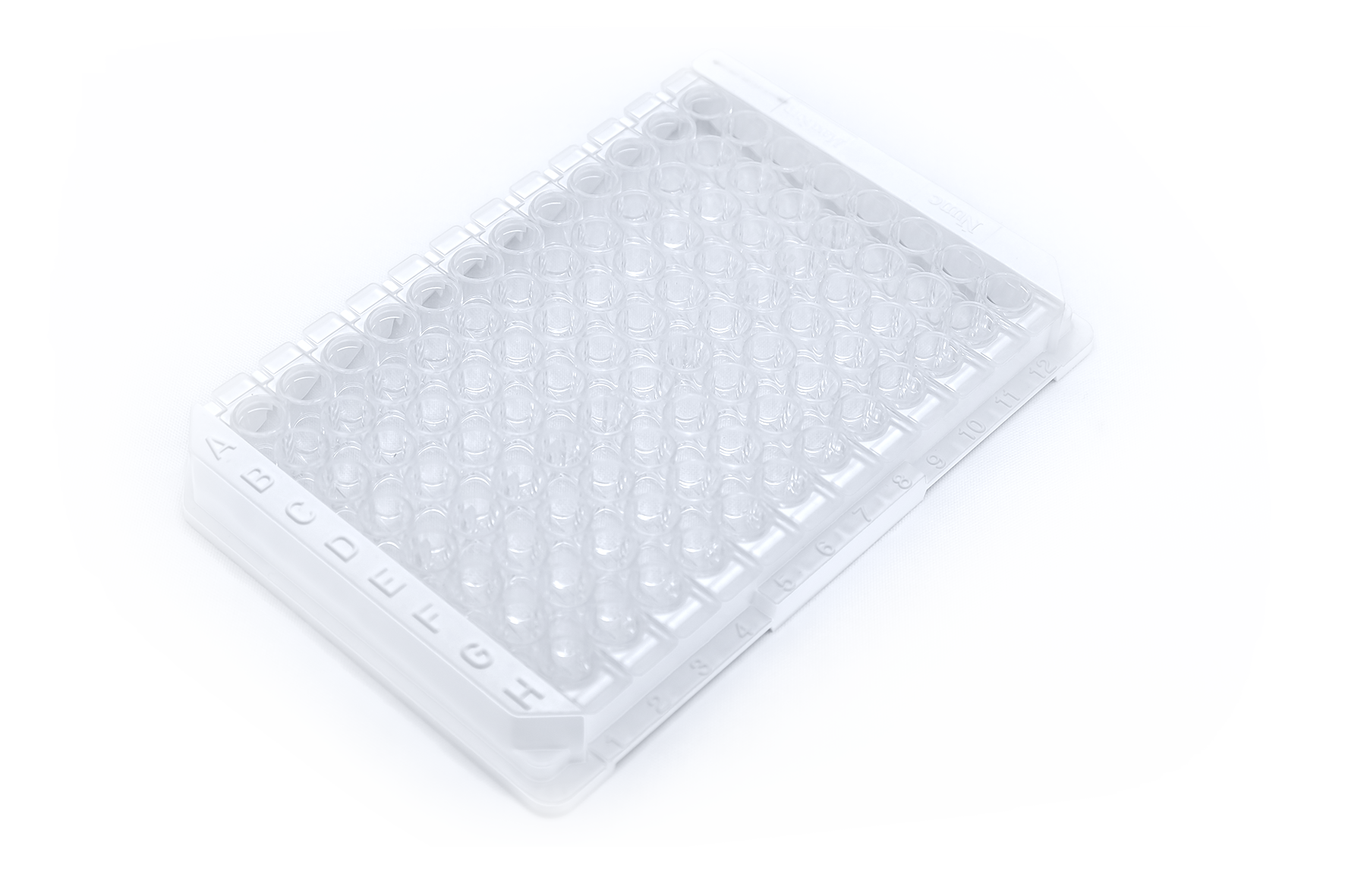
Allergens are bound on the surface of each well of the microtiter plate. For testing, a diluted sample (serum or plasma) is added to the wells. Specific IgE antibodies of the sample bind to the antigens in the wells during incubation, and non-bound components of serum or plasma are washed away after incubation. Subsequently, digoxigenin-labeled anti-human IgE antibodies and radish peroxidase coupled anti-digoxigenin antibodies (conjugate) are added.
The digoxigenin-labeled anti-human IgE antibodies bind to IgE antibodies of the sample, standards and controls. The conjugate binds to the digoxigenin-labeled anti-human IgE antibodies. Subsequently, un-bound detection antibodies are washed away. A color substrate TMB (3,3‘, 5,5‘-tetramethylbenzidine) is added, which is converted by the horseradish peroxidase conjugate bound enzyme. The reaction is stopped by adding a stop solution. A yellow dye is formed and the respective intensity correlates with the proportional amount of locally-bound antibody. The dye formed is quantified photometrically by measuring the extinction. The quantification of the tests is carried out using a point-to-point regression of a 6-point standard curve.
Standards are calibrated according to the WHO reference serum 75/5021. The identified units/mL can be assigned to the respective CAP-classes and provide the level of specific IgE sensitization (See table below).
| CAP-class | U/ml | Sensitisation |
| 0 | 0,00 - 0,34 | none |
| 1 | 0,35 - 0,69 | very low |
| 2 | 0,70 - 3,49 | low-moderate |
| 3 | 3,50 - 17,49 | moderate |
| 4 | 17,50 - 49,99 | high |
| 5 | 50,00 - 99,99 | very high |
| 6 | ≥ 100 | extremly high |
ELISA Kit for detection of total IgE

Test principle
The quantification of total IgE is based on a sandwich ELISA protocol (Enzyme Linked Immunosorbent Assay). The wells of the microtiter strips are coated with polyclonal anti-IgE antibodies which bind the existing immunoglobulin from the sample. Detection occurs with a HRP conjugate (horseradish), which makes the visual evaluation and quantification of bound antibodies possible. The results are analysed photometrically at 405 nm after the reaction is stopped.
| Age in years | U/ml |
| Newborns | < 2,0 |
| < 1 | 0 - 10,0 |
| 1 - 4 | 0 - 47,5 |
| 4 - 16 | 0 - 150,0 |
| > 16 | 0 - 120,0 |


